
MATERNAL-FETAL MEDICINE
Promising Results for in-Utero Treatment of Spina Bifida
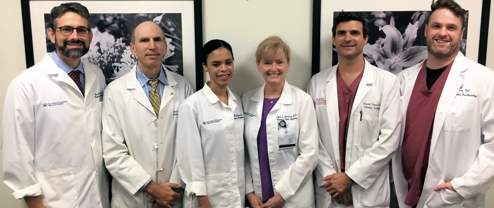
ver the past fifteen years, Dr. Lynn Simpson, Chief of the Division of Maternal Fetal Medicine, and Dr. Russ Miller, Medical Director of the Carmen and John Thain Center for Prenatal Pediatrics, have worked together to build a leading fetal diagnosis and therapy program at Columbia.
Through multidisciplinary collaborations with pediatric subspecialists, pediatric surgical specialists, genetics, anesthesia, and other departments across the medical center – and with the generous and visionary support of Carmen and John Thain – Drs. Simpson and Miller have collaborated to develop programs to treat many different types of fetal anomalies and conditions. One such example involves the use of fetoscopic laser photocoagulation to interrupt twin-twin transfusion syndrome (TTTS). The development of this laser program helped Columbia establish itself as a regional leader in fetal therapy and diagnosis, including the evaluation and management of complicated multiple gestations. Columbia is now widely regarded as a referral center for patients with complicated twin pregnancies, and Drs. Simpson and Miller are internationally recognized for their expertise in the management of complicated multiples.
Most recently, however, the team’s attention has also been focused on in-utero treatment of spina bifida, known as fetal myelomeningocele (fMMC) repair. This program involves a broad, successful multi-disciplinary collaboration that draws upon the expertise of numerous collaborators, including Dr. Vincent Duron, Columbia pediatric surgeon and program co-leader, Dr. Neil Feldstein, Columbia pediatric neurosurgeon, and Dr. Laurence Ring, Columbia obstetric anesthesiologist.
After a randomized controlled trial comparing open prenatal repair to postnatal repair demonstrated benefits, including a significantly reduced need for a ventriculoperitoneal (VP) shunt at 1-year post-birth and improved neuromotor outcomes at 2.5 years post-birth, many US centers began offering this type of prenatal surgery. However, prenatal repair is associated with significant maternal and obstetrical risks, including premature delivery, PPROM, bleeding, and uterine scar complications, as well as a need for cesarean delivery with all pregnancies. These potential issues must be considered by patients and families considering this operation.
In an effort to provide the benefits of prenatal surgery while limiting its maternal and obstetrical risk, the Columbia team uses a modified fetoscopic version of the technique that involves externalization of the uterus. While the technique still involves a major surgery for the mother, uterine access is achieved through the placement of small fetoscopic ports that are less morbid for patients, but achieve a similar fetal repair. In fact, patients who undergo this type of repair may even be candidates to undergo subsequent vaginal deliveries. Outcomes using this technique for fMMC repair have been extremely promising. Since 2019, the team has performed 13 cases, resulting in a 100% live birth rate, the option for vaginal delivery, and a VP shunt rate that is comparable to the published literature. The first 10 cases performed had a 36 1/7-week average gestational age at delivery.
“This technique significantly changes the outlook for families who receive an unexpected diagnosis during their pregnancies,” said Dr. Simpson. “The ability to not only diagnose prenatally, but treat prenatally – to move up the treatment window – reduces the severity of the lesion, minimizes the intensity of the treatment required, and improves overall outcomes for mother and baby.”